
Right Bronchus of the Lung
Anatomical Illustration (2019): unique view of the lungs, exposing the right bronchus created through observation of anatomical specimens (dissections), 3D models, and text references.
Media: Procreate, Horos, Zbrush
Process Work
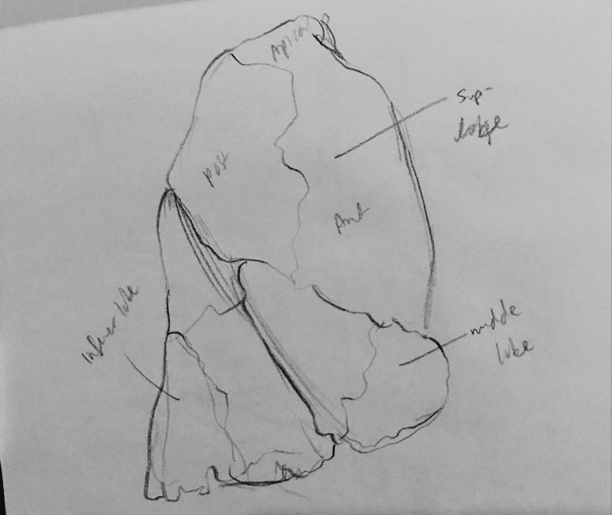
1 Research & Sketches
Sketches from observation were drawn to better understand the anatomy at different angles, and how the bronchus branched in 3D space based on preserved lungs and metal casts found in Grant’s Museum (University of Toronto). A rough 3D model was also created based on bilateral CT data of lungs using Horos and Zbrush.


2 Linear Drafts & Revisions
Multiple drafts of the line work were created and revised for anatomical accuracy and clarity.


3 Tonal Rendering & Color
A greyscale tonal render was first created to better understand the lighting. Color was then added as a separate layer.

