
Double-Lung Transplant: Anastomosis
Surgical Sequence Excerpt (2019): surgical sequence of the anastomosis of a donor lung created through live surgical observation and anatomical references.
Client: Dr. M Cypel, Dept of Thoracic Surgery, UHN
Media: Adobe Photoshop, Adobe InDesign
Process Work
1 Research


2 Surgical Observation
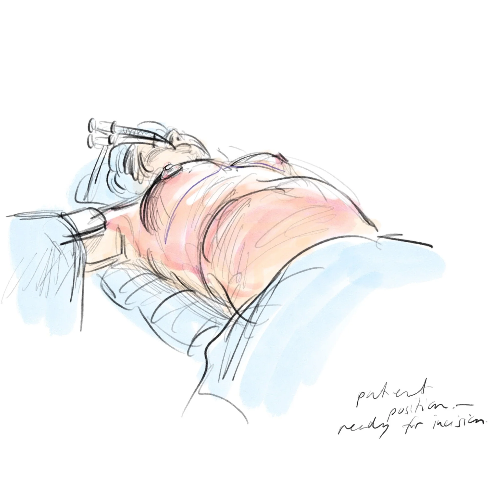
It was important to understand the procedure through observation and live study. This was done by observing two 10-hour double-lung transplant surgeries and multiple EVLP procedures. Sketches were done using a variety of mediums: pen, graphite, colored pencil, and ipad pro (using procreate).


3 Drafts & Revisions
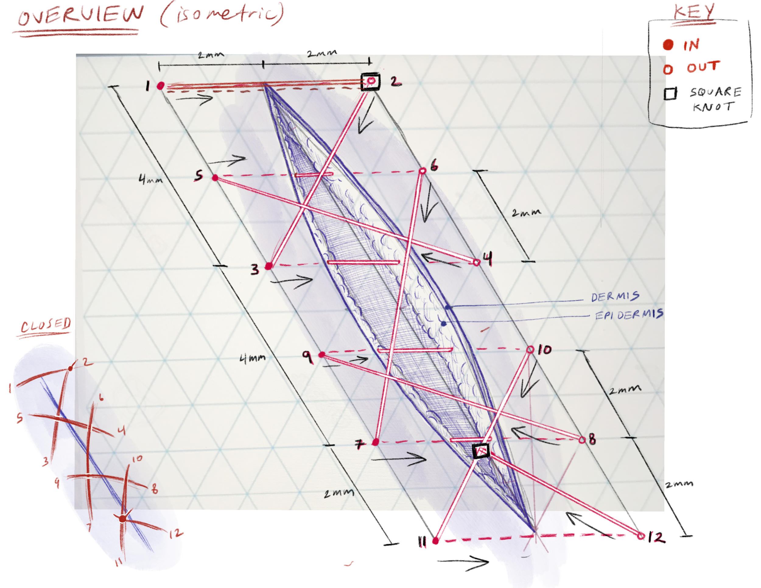
The sequence went through multiple drafts and revisions to correct anatomy, and visual placement of the elements for clarity. In real-life, the suturing technique illustrated is difficult to observe during surgery and through photographs. The illustrations needed to capture the steps in an accurate, but simplified manner.


4 Compositing
Final layout was created in Adobe InDesign.

